
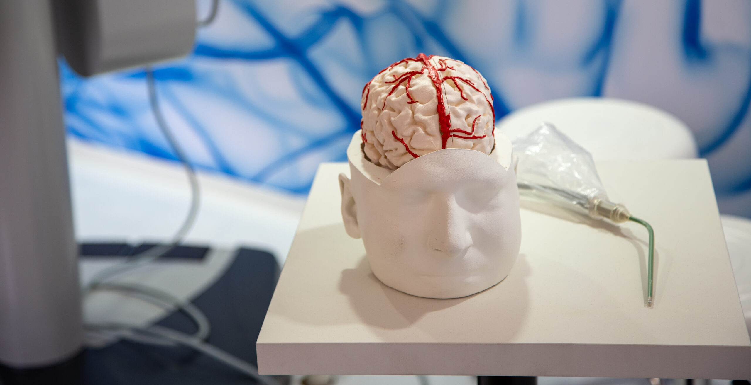
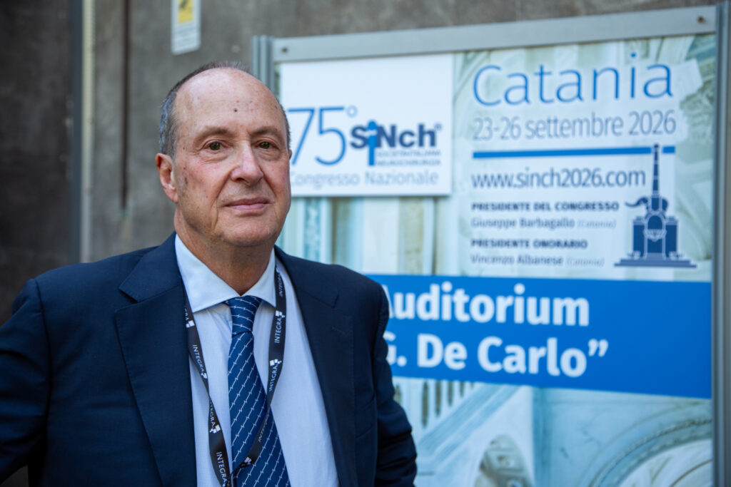
From September 23 to 26, the 75th National Congress of the Italian Society of Neurosurgery (SINch) was held in Catania. Among the more than 700 scientific contributions presented, several studies analyzed how artificial intelligence can support the neurosurgeon in interpreting diagnostic images, planning interventions, and evaluating prognosis.
The results are promising, but must be interpreted with caution. “Artificial intelligence can help the neurosurgeon assemble a volume of information that would be very difficult to examine manually within the same timeframe. It can point to elements deserving attention, but it does not know the patient’s history and does not make clinical decisions. The responsibility for choosing the course of action remains with the specialist,” says Domenico D’Avella, president of SINch.
Brain Tumors
Among the studies presented is an artificial intelligence system developed to analyze MRI scans of patients with glioma, validated on 1,251 cases. The model demonstrated superior performance compared with leading benchmarks, while also requiring less computational memory. The research focuses on gliomas, tumors that arise in the central nervous system and demand precise identification of the tumor’s distinct components in diagnostic images. This phase is essential for planning the surgical intervention, assessing its effectiveness, and monitoring disease progression.
A group of researchers developed the UNETR-RF system, capable of assessing the reliability of different MRI images and assigning greater weight to those deemed more informative, thereby reducing the impact of patient motion and differences between imaging devices. The researchers view these results as a first step toward the development of GliomAId, a platform designed to automatically distinguish tumor, surgical cavity, and any residual tissue. The system, however, remains in the research stage.
“Understanding precisely where the tumor lies and how it relates to surrounding structures is essential for preparing the operation. Systems like these could make image analysis faster and more uniform. Before clinical use, they must be rigorously validated and tested in different healthcare settings,” explains Giuseppe Barbagallo, president of the Congress and SINch Secretary.

Brain Connectivity
A second research project aims to help the neurosurgeon visualize and interpret the brain connections involved in movement, language, and key cognitive functions. The model developed by the researchers can organize large amounts of data obtained from tractography imaging, a technique that enables reconstructing the pathways of brain nerve fibers. The goal is to aid the surgeon in identifying neural tracts to preserve during surgery, even when tumors or other lesions alter brain anatomy.
“Understanding the organization of brain networks with increasing accuracy means being able to plan more personalized interventions. These are tools that could help us integrate vast amounts of information while keeping patient safety at the center,” notes Barbagallo.
AI Applied to Parkinson’s
Artificial intelligence could help make planning for deep brain stimulation more precise, a procedure used in some Parkinson’s patients to control symptoms such as tremor, rigidity, and slowness of movement. A study presented at the Congress evaluated a system capable of automatically identifying the anatomical target and proposing the electrode trajectory based on the patient’s brain images. The method was retrospectively validated on 10 patients and 20 trajectories. Automated processing took about 10 minutes and demonstrated greater accuracy than the conventional planning used as a comparator.
“The objective is not to replace the neurosurgeon, but to provide an additional tool to plan the procedure in a more personalized and accurate way. Looking ahead, technologies of this kind could help speed up image analysis and support more complex clinical decisions,” commented D’Avella.
From Aneurysm Prognosis to Risk Assessment
A study conducted on 326 patients treated across nine Italian centers examined brain hemorrhages caused by aneurysm rupture. The analysis identified the volume of blood present in the ventricles as the main factor associated with mortality and disability in the population studied. The study used artificial intelligence models to analyze the interaction between intraventricular hemorrhage volume, age, and other clinical parameters, with the aim of pinpointing the factors most strongly linked to patient outcomes.
Technology Does Not Replace the Doctor-Patient Relationship
Alongside the opportunities, there are concerns about protecting health data, the risk of errors, and the transparency of the systems used. An algorithm trained on limited populations might not maintain the same performance in patients with different characteristics. Moreover, results must be interpretable and critically evaluated by specialists, avoiding AI becoming a “black box” that automatically makes decisions.
As SINch experts emphasize, artificial intelligence represents an increasingly important support tool, but clinical judgment, the evaluation of the individual patient, and the physician-patient relationship remain central to the decision-making process.

Abbonati a Karla Miller