
Epigenetic changes are those that influence gene activity or expression without altering the DNA sequence in any way. Epigenetic modifications or marks occur on both histones and DNA through a network of proteins that induce changes, adding epigenetic marks (writers) and alter the existing state by removing marks or responding to specific epigenetic marks (readers).
These biochemical signals are transmitted to daughter cells during cell division (mitosis) and, in certain cases, to future generations through the gametes (meiosis). Environmental factors (such as diet, stress, physical activity, and pollution) can effectively influence the activity of genes, “turning them on” or “off,” without changing the DNA sequence. There are several broad groups of epigenetic modifications: acetylation, methylation, phosphorylation, and ubiquitination.
Specifically, in this discussion we address methylation, a process that modifies the genome without altering its sequence and enables rapid and lasting adaptation to environmental changes. Methylation involves the addition of a methyl group (-CH3) directly to DNA (primarily at a cytosine base) or to the tails of histones on lysine and arginine amino acids. It is useful to recall that histones are positively charged basic proteins that act as a “spool” for the negatively charged DNA. While DNA methylation is generally associated with gene silencing, because, when present in a promoter, it inhibits transcription and thus plays a role in cancer prevention. The introduction of methyl groups onto histone tails is associated with both gene activation and silencing, because, depending on the number of groups added and the specific amino acid target on the histone tail (e.g., lysine or arginine), it can compact the chromatin into heterochromatin and render it inaccessible to transcription factors, or methylation of other residues can have the opposite effect, relaxing the chromatin structure (euchromatin) around the gene and promoting transcription.
DNA methylation refers to the process whereby the 5′ end in the genomic DNA sequence is converted from cytosine to 5′-methylcytosine (5mC) by the action of a class of enzymes known as DNA methyltransferases. This process occurs primarily at CpG dinucleotides (a cytosine followed by a guanine), which often cluster in regions known as CpG islands. This epigenetic modification, while not altering the gene sequence, can influence the pattern of gene expression and represents one of the main forms of epigenetic regulation of gene expression in mammals.
As an important epigenetic modification, DNA methylation plays a crucial role in modern biomedical research. CpG islands are genomic regions in which CpG sequences are tenfold more frequent and are often located near transcription start sites.
As mentioned above, the enzymes responsible for methylation are a specific class known as DNA methyltransferases (DNMTs). In particular, DNMT1, a member of this family, is expressed in proliferating cells and is responsible for propagating methylation patterns in the daughter DNA strands during replication. Although its expression is reduced in adult somatic tissue, methylation is prominent during early embryonic development and is thought to be carried out by the isoenzymes of the family referred to as DNMT3A and DNMT3B at regions deprived of nucleosomes in the genome. Another isozyme of the family, DNMT3L, helps target and regulate the function of DNMT3A/3B and plays a role in de novo methylation; it is effectively a non-catalytic homolog of DNMT3A/B that aids their binding to the target DNA.
Additional proteins and histone deacetylases (HDACs) help direct DNMT proteins to the appropriate regions of the genome, regulate methyltransferase activity, and coordinate further epigenetic marks with DNA methylation.
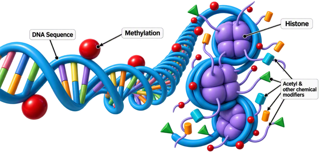
In summary, we can thus define two types of DNA methylation:
- Maintenance methylation: describes the process by which a already methylated DNA duplex guides the modification of the corresponding unmethylated strand during DNA replication, thereby preserving the original methylation pattern. This form of methylation is primarily catalyzed by maintenance methyltransferase (such as DNMT1), which accurately copies the patterns established on the parental DNA strands to the daughter strands during replication, ensuring stable transmission of epigenetic information. Clinically, it has significance for research on inherited diseases.
- De novo methylation: refers to the process of adding methyl groups to newly formed DNA sites without relying on existing methylated DNA strands. This form of methylation occurs mainly during early embryonic development and in some cellular reprogramming processes. It involves specific methyltransferases, such as DNMT3A and DNMT3B, which can add methyl groups to DNA molecules in the absence of a preexisting methylation pattern. Clinically, this mechanism is applicable to studies of embryonic development, cellular differentiation, and cancer research.
So, what is the purpose of DNA methylation? It serves two crucial functions:
- gene expression inhibition because cytosine methylation is tightly linked to the formation of “silent” chromatin;
- maintenance of genomic integrity through the silencing of parasitic sequences: methylation is essential to keeping genomes of transposons and retrotransposons in a silent state.
Hypo- and hypermethylation of DNA: correlation with carcinogenesis
DNA hypomethylation is an epigenetic modification characterized by a reduction of methyl groups on the genome, leading to activation or overexpression of genes. This process is fundamental for development, but when abnormal it can cause chromosomal instability, accelerated biological aging, and promote tumor formation. As described, DNA methylation generally acts as a “stop switch” to silence genes. When hypomethylation occurs, this block is removed.
Typically, CpG islands (regions where cytosine-guanine nucleotides occur in tandem at a frequency >50%) in normal cells are hypermethylated relative to the rest of the genome to silence certain genes.
However, if CpG islands within the promoter regions of tumor suppressor genes (TSGs) become hypermethylated by DNMTs, which are typically overexpressed in cancer, tumorigenicity increases by reducing the cell’s ability to suppress tumors. Hypermethylation of CpG islands can disrupt binding to the CTCF protein (CCCTC-binding factor), leading to loss of key insulating regions and consequent oncogene activation; this is a protein that serves as a scaffold in mediating genome organization and gene expression within it.
All the DNA methyltransferases mentioned earlier—DNMT1, DNMT3A, and DNMT3B—are overexpressed in many cancers, including acute and chronic myeloid leukemias, as well as colon, breast, and stomach carcinomas. Hypermethylation of gene bodies and intergenic regions of DNA is also linked and can contribute to cancer development.
DNA methylation erasers
DNA methylation becomes pathological when CpG islands within oncogene regulatory regions are affected, such as TP53, CDKN2A (p16), BRCA1, BRCA2, and RB1, to name a few key examples; 5-methylcytosine, once viewed as a passive mark left behind during DNA replication and established de novo by DNMT3A and DNMT3B and maintained by DNMT1, was originally thought to be passively exhausted during replication. However, subsequent studies have shown that active removal is mediated by a special family of enzymes called the TETs (Ten-Eleven Translocation), which catalyze the oxidation of methylated cytosine (5mC) to 5-hydroxymethylcytosine (5hmC) and then to other oxidized forms, enabling active DNA demethylation. This occurs specifically through sequential oxidation mediated by TET1, TET2, or TET3 of 5-methylcytosine (5-mC) to 5-hydroxymethylcytosine (5-hmC), 5-formylcytosine (5-fC), and 5-carboxylcytosine (5-caC). This iterative oxidation is favored by the presence of oxygen, as well as bivalent iron ions and 2-oxoglutarate–dependent enzymes.
Moreover, CpG island demethylation allows the CTCF protein to bind DNA, positively regulating gene activation and insulating functions performed by this protein, as it prevents aberrant interactions between promoters and enhancers, promoting proper three-dimensional chromatin organization.
The TETs play a fundamental protective role: loss or reduction of their activity (via mutations or inhibition by oncometabolites) leads to a DNA hypermethylation phenotype, with silencing of tumor suppressor genes and transcriptional misregulation, associated with aberrant hypermethylation, genomic instability, and cancer development. While lower levels of 5hmC (the direct product of TET activity) are a negative epigenetic marker that correlate with tumor progression and unfavorable prognosis in several cancers, 5hmC has been proposed as an early biomarker for diagnosis and monitoring of neoplasms, thanks to ultrasensitive new technologies. To cite an example, a recent meta-analysis of 1,736 cancer patients shows that low levels of 5hmC are associated with an increased risk of lymph node metastasis and advanced disease stage, as well as significantly poorer overall survival.n
It should be emphasized that TETs also protect genomic stability through the regulation of retrotransposons and by preventing derepression of transposable elements, which can lead to genomic damage and developmental disruptions.
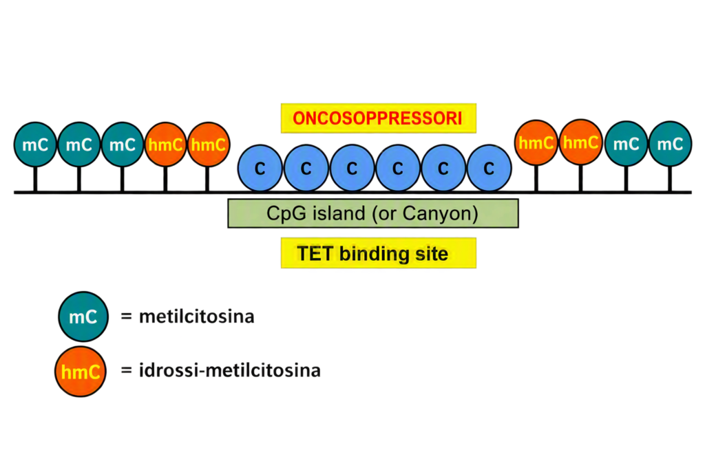
Figure 2 illustrates the TET action sites where, in the presence of oxygen, the hydroxylation of methylcytosine acts as a protective cushion to prevent hypermethylation at CpG islands near tumor-suppressor genes.
The figure shows the distribution of methyl groups along a DNA strand:
- CpG Islands / Canyon: central unmethylated zones (blue spheres with the C).
- Edges: presence of hmC (red spheres), indicating ongoing demethylation activity.
- Outer regions: presence of mC (purple spheres), representing methylated and silenced DNA.
Cellular hypoxia as a driver of tumorigenesis
Cellular hypoxia alters DNA methylation via suppressed epigenome oxidation. The conversion of 5-methylcytosine to 5-hydroxymethylcytosine by the TET enzymes requires oxygen, while hypoxia is common in cancer, suggesting a potential synergy. Furthermore, hypoxia induces the activity of hypoxia-inducible transcription factors (HIFs), which alter gene expression in cells to cope with low oxygen and can promote a metabolic shift from oxidative phosphorylation to anaerobic glycolysis, an event that can drive the Warburg effect exploited by tumor cells to survive under hypoxic conditions.
As described above, hypermethylation of tumor suppressor gene promoters (TSGs) provides growth advantages to cancer cells, but how these changes present themselves has been poorly understood until now.
With the CHAMELEO study (named because it was conducted on chameleon cells), it is shown that tumor hypoxia reduces the activity of oxygen-dependent TET enzymes, which catalyze DNA demethylation through the oxidation of 5-methylcytosine.
The loss of TET activity depended solely on oxygen scarcity and increased hypermethylation of gene promoters. Similar observations on promoter methylation have been made in tumor samples from patients where genes involved in DNA repair, cell cycle regulation, angiogenesis, and metastasis are frequently implicated. This also suggests clonal selection of hypermethylation events that favor growth and metastatic spread.
Overall, the CHAMELEO study confirms the tumor hypoxia–mediated regulatory role on DNA methylation and, importantly, suggests that strategies aimed at normalizing intratumoral oxygen levels could reverse epigenetic changes and potentially modulate tumor cell properties. Thus, tumor hypoxia acts as a new regulator at the base of DNA methylation. This emphasizes the importance of maintaining proper cellular oxygenation to prevent the initiation of pathogenetic mechanisms that can cause genomic instability and activation of oncogenic genes.
Vitamin C or ascorbate supplementation can boost TET activity and DNA demethylation in embryonic stem cells, suggesting a possible environmental regulatory role, while retinol vitamin A supplementation appears capable of increasing TET expression.
Regarding natural substances, several recent studies suggest they can curb the aberrant action of DNA methyltransferases expressed in tumor cells; these include green tea catechins, particularly epigallocatechin gallate (EGCG), broccoli-derived sulforaphane, resveratrol, quercetin, and finally selenium.
In summary, the TET enzymes are key players in protecting epigenetic and genomic integrity, counteracting aberrant DNA methylation and contributing to cancer prevention and immune regulation.
The nucleosome
The nucleosome is the fundamental structural and functional unit of chromatin and consists of a segment of about 147 base pairs of DNA wrapped almost twice around an octamer of histone proteins (two molecules each of H2A, H2B, H3, and H4). The nucleosomes are responsible for the first level of genetic material compaction in the cell nucleus and are often described as “beads on a string.” This organization allows the long DNA strand to fit within the microscopic space of the cell and regulate gene expression. Beyond packaging DNA, nucleosomes play a key role in regulating gene expression because the histone tails protrude from the protein complex and can undergo chemical modifications, determining whether a particular region of DNA is accessible for transcription.
Histone acetylation
Histone acetylation is another epigenetic modification in which enzymes called HATs (Histone Acetyltransferases) add acetyl groups to the amino acid tails of histones, reducing the positive charge of the proteins and loosening their grip on DNA. The result is a more open chromatin structure that allows transcription factors to access DNA and activate gene expression. The process involves two main classes of enzymes acting in opposition:
- Histone Acetyltransferases (HATs): add acetyl groups, promoting chromatin relaxation and gene activation.
- Histone Deacetylases (HDACs): remove acetyl groups, compacting chromatin and repressing transcription.
Interestingly, HDACs are divided into several classes that include the various sirtuins, a topic addressed in a previous issue of the journal (September 2026).
The balance between HAT and HDAC activity is modulated and sensitive to cellular signals, the environment, and the state of cellular differentiation. Disturbances in this “epigenetic balance” can contribute to pathological conditions, including cancer, neurodegenerative diseases, and metabolic disorders.
Transposons
Transposons are segments of DNA capable of moving within the genome, causing mutations such as deletions, inversions, and translocations. Retrotransposons are transposons that are transcribed into RNA and subsequently into DNA, using RNA as a transport mechanism similar to retroviruses. The enzyme involved is reverse transcriptase. In molecular biology, this enzyme is the fundamental tool behind the RT-PCR (Reverse Transcription Polymerase Chain Reaction), used to detect and amplify RNA sequences and widely employed in biomedical laboratories for molecular testing.
Adapted from the October 2026 issue of Karla Miller
Bibliography
- Bestor TH. The DNA methyltransferases of mammals. Hum Mol Genet. 2000; 9:2395–402.
- Jurkowska RZ, Jeltsch A. Enzymology of Mammalian DNA Methyltransferases. Adv Exp Med Biol. 2016;945:87-122.
- DiTroia SP, Percharde M, Guerquin MJ, Wall E, Collignon E, et al. Maternal vitamin C regulates reprogramming of DNA methylation and germline development. Nature. 2019; 573:271–5.
- Ito S, D’Alessio AC, Taranova OV, Hong K, Sowers LC, Zhang Y. Role of TET proteins in 5mC to 5hmC conversion, ES-cell self-renewal and inner cell mass specification. Nature. 2010; 466:1129–33
- Kim MS, Lee J, Sidransky D. DNA methylation markers in colorectal cancer. Cancer Metastasis Rev. 2010; 29:181–206
- Wu X, Zhang Y. TET-mediated active DNA demethylation: mechanism, function and beyond. Nat Rev Genet. 2017;18(9):517-534.
- Ginder GD, Williams DC. Readers of DNA Methylation, the MBD family as potential therapeutic targets. Pharmacology & Therapeutics. 2018; 184:98-111.
- Bergman Y, Cedar H. DNA methylation dynamics in health and disease. Nat Struct Mol Biol. 2013;20(3):274-81.
- Shi DQ, Ali I, Tang J, Yang WC. New Insights into 5hmC DNA Modification: Generation, Distribution and Function. Front Genet. 2017; 8:100.
- Clouaire T, Stancheva I. Methyl-CpG binding proteins: specialized transcriptional repressors or structural components of chromatin?. Cell Mol Life Sci. 2008;65(10):1509-22.

Abbonati a Karla Miller