
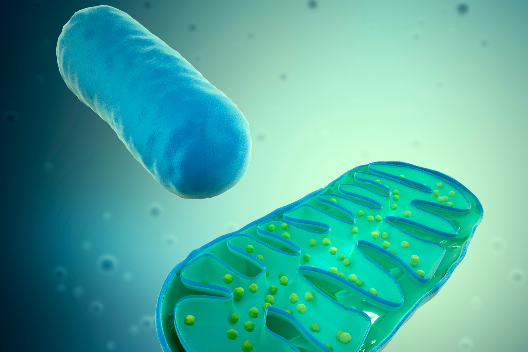
Mitochondria originated from alpha-proteobacterial endosymbionts and evolved from ancestral bacterial progenitors through symbiosis within host eukaryotic cells. They are characterized by a double-membrane structure, consisting of an outer membrane and an inner membrane that encloses the intermembrane space (IMS) and the mitochondrial matrix.
The inner membrane forms cristae and houses numerous enzymes and protein complexes. Beneath the inner membrane lies the mitochondrial matrix, which contains a circular genome known as mtDNA and multiple copies of ribosomes. Unlike nuclear DNA (nDNA), mtDNA generally lacks introns and replicates independently of the host genome.
The human mtDNA is about 16,569 base pairs long and encodes 37 genes, whereas nuclear DNA is thought to contain between 20,000 and 25,000 genes. It is not organized into chromosomes, while nuclear DNA is partitioned into 23 chromosomes and, with certain proteins, forms a substance called chromatin. Finally, it includes a series of nucleotides that participate in two genes simultaneously, whereas nuclear DNA has genes whose nucleotide sequences are well defined and distinct from one another.
In virtue of the mitochondrial origin mentioned above, mitochondrial DNA most likely has a bacterial origin. In fact, based on numerous independent studies, molecular biologists believe that the presence of mitochondrial DNA in cells results from the incorporation, by ancestral eukaryotic cells, of independent bacterial organisms, very similar to mitochondria.
This discovery has surprised only partly the scientific community, as DNA found in bacteria is generally a circular strand of nucleotides, exactly like mtDNA. The widely accepted theory that mitochondria and mitochondrial DNA have a bacterial origin is known as the “endosymbiotic theory,” where “endosymbiosis” denotes a collaboration between two organisms that involves engulfing one within the other to gain a certain advantage. The 37 genes of mitochondrial DNA encode: proteins, transfer RNA (tRNA) and ribosomal RNA (rRNA). Specifically:
- 13 encode for 13 proteins that constitute the enzymatic subunits of the mitochondrial respiratory chain dedicated to oxidative phosphorylation;
- 22 encode for 22 tRNA molecules;
- 2 encode for 2 rRNA molecules.
The tRNA and rRNA molecules are fundamental to the synthesis of the aforementioned 13 proteins, as they form the machinery that governs their production.
Therefore, in other words, mitochondrial DNA holds the information to produce a definite set of proteins and the tools necessary for their synthesis.
The 13 protein-coding genes are organized into two main clusters: one encodes the subunits of Complex I (NADH dehydrogenase), while the other encodes the subunits of the remaining respiratory complexes, specifically Complex III (cytochrome bc1), Complex IV (cytochrome c oxidase) and Complex V (ATP synthase). These proteins are essential components of the electron transport chain during oxidative phosphorylation.
The mtDNA also contains several non-coding regions and, in particular, a control region within which lies the D-loop (displacement loop) that forms when a short DNA strand (called 7S DNA) inserts stably into the double helix, separating the two original strands, and creating a looped structure that contains the promoter of the heavy strand (HSP1 and HSP2) and a promoter for the light strand (LSP), acting as a fundamental control hub for gene expression and the origin of replication of the heavy strand. This unique structure provides the physical and chemical environment necessary for mitochondria to perform their diverse functions.
The role of mitochondria in bioenergetics, homeostasis, and cellular signaling
Recent discoveries have shown that mitochondria play several central roles in cells beyond their primary energy function. Mitochondria are now recognized as essential organelles not only for energy conversion but also for signal transduction and regulation of cell death. The principal function of mitochondria remains the generation of ATP through oxidative phosphorylation (OXPHOS). In this process, cells extract electrons from the nicotinamide adenine dinucleotide (NADH) and from the flavin adenine dinucleotide (FADH2), biologically active forms of vitamins B3 and B2 respectively, and use them to pump protons across the inner mitochondrial membrane (IMM), creating a proton gradient. This gradient drives ATP synthesis via the ATP synthase, a enzymatic complex embedded in the IMM, which effectively constitutes Complex V of the respiratory chain, whose functional understanding clarified, in a major way, the mechanism of mitochondrial energetics and relies on the chemiosmotic hypothesis proposed by the British biochemist Peter Mitchell in 1961, a theory that earned him the Nobel Prize in Chemistry in 1978.
Electrons originating from NADH and FADH2 are transferred through a series of protein complexes in the electron transport chain (ETC), ultimately combining with oxygen to form water—a process that is coupled to the generation of the proton-motive force. The mitochondrial respiratory chain (MRC) is a key energy converter in eukaryotic cells, comprising four complexes (I–IV) and two electron carriers embedded in the IMM.
The four complexes transfer electrons from various metabolic sources to molecular oxygen, establishing an electrochemical gradient across both sides of the IMM to drive ATP synthesis. Complex I catalyzes the oxidation of reduced NADH and the reduction of ubiquinone (the oxidized form of Coenzyme Q10), with two electrons and four protons contributed, promoting the formation of a proton gradient across the IMM.
Complex II oxidizes succinate to fumarate and reduces ubiquinone to ubiquinol, the reduced form of Coenzyme Q10, for electron transfer. Complex III couples the oxidation of ubiquinol to the reduction of cytochrome c: during electron transfer, it absorbs two protons from the mitochondrial matrix and releases four into the intermembrane space. The release of 4 protons into the intermembrane space generates a proton-motive force essential for ATP production via ATP synthase. Complex IV transfers the electrons that cytochrome c donates directly to oxygen, reducing it to water. In this process, two protons are moved against the concentration gradient.
The Complex V, namely the aforementioned ATP synthase, comprises two functional domains, F0 and F1, which use the created proton gradient to catalyze ATP synthesis, via a mechanism that can be likened to a water-wheel turning energy. This occurs because protons accumulated in the intermembrane space cannot re-enter the matrix because the inner mitochondrial membrane is impermeable to H+; thus they must pass through the ATP synthase channel: the proton flow through this specialized protein drives ATP production that underpins cellular energetics.
Note the central role of Coenzyme Q10, in its oxidized and reduced forms, as a true electron shuttle intercalated among complexes I, II, and III.
Mechanisms of mitochondrial signal transduction are complex and diverse, covering a wide range of intracellular-to-extracellular signaling recognition and processing, including oxidative stress, the mtROS signaling pathway which represents the mechanism by which reactive oxygen species produced by mitochondria act as second messengers, Ca2+ signaling, signaling of the mitochondrial genome, thermal signaling, and inflammatory signaling. Voltage-dependent anion channels (VDAC) in the outer mitochondrial membrane mediate signal transduction between the cytosol and the mitochondria. In addition, mitochondrial dynamics, including movement, fission and fusion—which we will discuss shortly—are closely linked to these signaling pathways.
Mit mitochondria work in concert with the nucleus and numerous organelles to build a system of mitochondrial information processing (MIPS), which can be summarized in three links. First, mitochondria have the capacity to perceive signals: these signals include changes in metabolite concentrations, hormonal signals, and oxidative stress. Second, the integration of information and processing signals occurs through physical interactions and diffusion mechanisms. Finally, mitochondria produce output signals that regulate the function of other organelles and systemic physiology. These output signals can influence cellular metabolism, gene expression, the cell cycle, and other aspects, thereby regulating cellular physiological activities.
Mitochondrial dynamics
Mitochondrial dynamics refer to the ongoing, dynamic process of fission and fusion within the cell, opposing but balanced processes that regulate the distribution, quantity, size, and shape of mitochondria in a dynamic equilibrium. Specifically, it includes, beyond fusion and fission, mitophagy and mitochondrial transport: a critical regulatory mechanism that maintains mitochondrial integrity and metabolic homeostasis inside the cells.
Mitochondrial fission and fusion
Scientific evidence indicates that mitochondrial fission and fusion are dynamic processes regulated by specific proteins that control mitochondrial morphology and function. Mitochondrial fission is the process by which a single mitochondrion divides into two or more distinct organelles and is mainly mediated by the cytosolic GTPase dynamin-related protein 1 (Drp1), which associates with the outer mitochondrial membrane via specific adaptors to form the “mitochondrial divisome” to execute division.
Drp1 interacts with four outer membrane–binding proteins, including the mitochondrial fission protein 1 (Fis1), the fission factor (Mff), the mitochondrial dynamics proteins MiD49 and MiD51. Drp1 and Fis1 are nonetheless the most critical proteins involved in mitochondrial division. Drp1 is recruited to sites of mitochondrial constriction, often facilitated by interactions with actin and the endoplasmic reticulum (ER), leading to the formation of two daughter mitochondria. It is an essential process for biogenesis (the creation of new mitochondria during cell division) and for the response to stress. Fission isolates heavily damaged portions of mitochondria or those with mutated DNA to allow their destruction through selective degradation (mitophagy), safeguarding the rest of the network.
In contrast, mitochondrial fusion is the process by which two or more mitochondria join to form a continuous tubular network. It involves two classes of dynamin-like proteins: the mitofusin 1 (Mfn1) and the mitofusin 2 (Mfn2), which mediate outer membrane fusion, while inner membrane fusion is regulated by the optic atrophy 1 (OPA1) protein. The process allows mitochondria to mix their contents (proteins, lipids, and mitochondrial DNA – mtDNA); in this way, healthy mitochondria can “compensate” those that are partially damaged by sharing intact genetic material, maximizing energy production efficiency (ATP).
These processes are regulated by complex mechanisms that include protein–protein interactions, post-translational modifications, and signaling responses to stress and metabolic needs. The presence of both mitofusins Mfn1 and Mfn2 markedly increases fusion efficiency. In particular, lack of Mfn1 leads to pronounced mitochondrial fragmentation, while deficiency of Mfn2 results in a higher proportion of mitochondria with spherical or ovoid shapes.
The coordinated regulation of fission and fusion is crucial to maintain mitochondrial quality, the distribution of mtDNA, and cellular energy function.
The perfect balance between fission and fusion is the key to the cell’s metabolic adaptation. If these mechanisms are disrupted, serious pathologies can arise. A shift too far toward fission fragments mitochondria and can lead to apoptosis (programmed cell death), while a defect in fission prevents the removal of damaged mitochondria. Dysfunctions in these processes are implicated in neurodegenerative diseases (e.g., Parkinson’s), cardiac conditions, and various forms of cancer.
Mitophagy and mitochondrial quality control
Mitophagy, the selective autophagy of mitochondria, is a process that directs damaged or dysfunctional mitochondria toward degradation, playing a fundamental role in mitochondrial quality control. When mitochondria are damaged or during ischemia or hypoxia, mutations in mitochondrial DNA (mtDNA) accumulate gradually under the influence of reactive oxygen species (ROS), leading to a reduction in the intracellular mitochondrial membrane potential (Δψm) and depolarizing damage, which triggers mitophagy.
The molecular mechanisms of mitophagy include ubiquitin-dependent pathways, specifically the “PINK1–Parkin pathway,” and ubiquitin-independent receptor-mediated pathways. In either case, both pathways culminate in the encapsulation of mitochondria within autophagosomes, double-membrane vesicles that the cell uses to engulf and isolate damaged or no longer needed components, followed by destruction through fusion with lysosomes in the cytoplasm.
PINK1-Parkin pathway
When a mitochondrion is damaged, the loss of mitochondrial membrane potential leads to the accumulation of the kinase PINK1 on the outer membrane, which activates the ubiquitin ligase Parkin, ubiquitinating mitochondrial proteins and signaling them for recruitment of autophagy receptors and the formation of an autophagosome.
The ubiquitination of mitochondrial proteins is central for selective recognition by autophagy receptors, which facilitate the incorporation of damaged mitochondria into autophagosomes.
Mitochondrial receptors
Some receptors on the mitochondrial membrane possess in their cytosolic domain a specific peptide sequence called LIR (LC3-Interacting Region), which binds directly to LC3 molecules present on the forming autophagosome in response to hypoxic stress or other stimuli.
Elimination via lysosome
The formed autophagosomes fuse with lysosomes, degrading mitochondria and recycling their components. In brief, a physiological mitophagy maintains the healthy mitochondrial population, preventing ROS accumulation and cellular damage, while a pathological mitophagy can lead to alterations in the process that are associated with neurodegeneration, cardiovascular disease, cancer, renal fibrosis, and autoimmunity.
Mitochondrial biogenesis is the process by which the cell increases its mitochondrial mass, involving replication of DNA, transcription, translation, and transport of lipids between organelles. Mitochondrial retrograde signaling, such as ATP levels, ROS, and Ca2+, modulates intracellular signaling cascades that influence mitochondrial biogenesis.
It is regulated by signaling cascades triggered by physiological stress (e.g., exercise or caloric restriction) that activate the transcriptional coactivator PGC-1-α.
- Perception of the stimulus: metabolic signals (such as an increased AMP/ATP ratio) activate cellular kinases like the enzyme AMPK, or deacetylases such as sirtuins.
- Transcriptional activation: these kinases phosphorylate and activate PGC-1α, the main regulator of biogenesis.
- Protein synthesis: PGC-1-α binds and activates the nuclear respiratory factors NRF1 and NRF2, which promote transcription of genes for mitochondrial proteins. These proteins are then synthesized in the cytoplasm and imported into the organelles.
- Genome expansion: concurrently, the transcription factor TFAM is activated to replicate and transcribe mtDNA.
In summary, mitochondrial dynamics is a highly regulated process, fundamental to mitochondrial and cellular function. Understanding the molecular mechanisms governing this dynamics is crucial to clarifying how mitochondrial form interfaces with function and to deepening our knowledge of diseases linked to defects in mitochondrial morphology.
The ubiquitination is a post-translational modification in which the protein ubiquitin covalently attaches to a target protein. This “tagging” serves as a molecular signal, guiding the protein toward degradation to maintain cellular homeostasis or modulating other functions.
The transcriptional coactivator PGC-1α is a master regulator of energy metabolism and mitochondrial biogenesis. It stimulates the creation of new mitochondria, enhances ATP production, and modulates fatty acid and glucose oxidation. For these reasons, it is highly expressed in tissues with high energy demand (such as skeletal muscle, heart, and liver) and is activated by physiological stimuli like exercise and exposure to cold.
Article from the September 2026 issue of Karla Miller
Bibliography
- Anderson S, Bankier AT, Barrell BG, et al. Sequence and organization of the human mitochondrial genome. Nature. 1981;290(5806):457-465.
- Friedman JR, Nunnari J. Mitochondrial form and function. Nature. 2014;505(7483):335-343.
- Hatefi Y. The mitochondrial electron transport and oxidative phosphorylation system. Annu Rev Biochem. 1985;54:1015-1069.
- Galluzzi L, Kepp O, Kroemer G. Mitochondria: master regulators of danger signalling. Nat Rev Mol Cell Biol. 2012;13(12):780-788.
- Kalia R, Wang RY, Yusuf A, et al. Structural basis of mitochondrial receptor binding and constriction by DRP1. Nature. 2018;558(7710):401-405.
- Palmer CS, Osellame LD, Laine D, Koutsopoulos OS, Frazier AE, Ryan MT. MiD49 and MiD51, new components of the mitochondrial fission machinery. EMBO Rep. 2011;12(6):565-573.
- Friedman JR, Lackner LL, West M, DiBenedetto JR, Nunnari J, Voeltz GK. ER tubules mark sites of mitochondrial division. Science. 2011;334(6054):358-362.
- Losón OC, Song Z, Chen H, Chan DC. Fis1, Mff, MiD49, and MiD51 mediate Drp1 recruitment in mitochondrial fission. Mol Biol Cell. 2013;24(5):659-667.
- Vives-Bauza C, Zhou C, Huang Y, et al. PINK1-dependent recruitment of Parkin to mitochondria in mitophagy. Proc Natl Acad Sci U S A. 2010;107(1):378-383.
- Tilokani L, Nagashima S, Paupe V, Prudent J. Mitochondrial dynamics: overview of molecular mechanisms. Essays Biochem. 2018;62(3):341-360.
- Raynald Bergeron, Jian Ming Ren, Kevin S. Cadman, Irene K. Moore, Pascale Perret, Marc Pypaert, Lawrence H. Young, Clay F. Semenkovich, and Gerald I. Shulman. American Journal of Physiology-Endocrinology and Metabolism 2001 281:6, E1340-E1346
- Fan H, Ding R, Liu W, et al. Heat shock protein 22 modulates NRF1/TFAM-dependent mitochondrial biogenesis and DRP1-sparked mitochondrial apoptosis through AMPK-PGC1α signaling pathway to alleviate the early brain injury of subarachnoid hemorrhage in rats. Redox Biol. 2021;40:101856.
- Toyama EQ, Herzig S, Courchet J, et al. Metabolism. AMP-activated protein kinase mediates mitochondrial fission in response to energy stress. Science. 2016;351(6270):275-281.
- Laker RC, Drake JC, Wilson RJ, et al. AMPK phosphorylation of Ulk1 is required for targeting of mitochondria to lysosomes in exercise-induced mitophagy. Nat Commun. 2017;8(1):548.
Abbonati a Karla Miller